
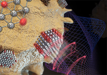
A technique known as super-resolved photonic force microscopy exploits optical trapping and artificial intelligence to detect exceptionally weak forces acting on nanoparticles in liquids. [Image: Lei Ding]
Scientists in China and Australia have shown how optically trapped nanoparticles can be used to measure miniscule forces in water at high spatial resolution (Nat. Photonics, doi: 10.1038/s41566-024-01462-7). They say that their new scheme, which relies on optical astigmatism and the pattern-spotting ability of neural networks, might shed light on protein function and improve disease diagnosis.
Measuring tiny forces
Many biological phenomena involve exceptionally small forces; both the tension in a DNA molecule and the effort needed to organize genetic material in a chromosome have magnitudes of less than 10-12 newtons. Researchers have already developed a number of techniques to measure such forces, but all have their weak points. Microscopy that relies on tiny cantilever arms, for example, is sensitive at such levels but struggles to map out the distribution of forces in three dimensions.
One particularly promising technique is photonic force microscopy. This involves holding a tiny dielectric particle in place using optical tweezers and collecting light scattered off the particle. Interference between scattered and unscattered light reveals the particle's three-dimensional position and with that, any forces acting on the particle.
Quantification of forces in a liquid has to account for a particle's buffeting by the liquid's molecules, and relies on measuring a shift in the distribution center of the particle's fluctuating position. Because the sampling rate of such measurements is limited by the autocorrelation time―how long it takes a particle to feel the trapping potential―this process imposes a thermal limit on force sensing.
To date, this nanoscale thermal limit has remained out of bounds. The best reported sensitivity for force measurements in solution is 10 femtonewtons (fN) per square root of the bandwidth (10 fN Hz–1/2; 1 fN = 10-15 N), achieved with a particle 500 nm in diameter.
These particles can be trapped via a phenomenon known as ion resonance, using a power of only around 10 milliwatts and causing a temperature rise of just 0.7°C.
Using smaller particles could reduce the drag experienced in a liquid―in principle raising measurement sensitivity―but doing so comes up against a couple of major hurdles. One is the lower scattering signal generated. The other is the need to increase trapping power due to lower trapping efficiency, which produces unwanted heat—oft-used gold nanoparticles are associated with a roughly 40°C temperature rise that would damage biological samples.
Measurements in three dimensions
In the latest work, Fan Wang, Beihang University, China, and colleagues show how to overcome these problems using probes in the form of lanthanide-doped nanoparticles. These particles can be trapped via a phenomenon known as ion resonance, using a power of only around 10 milliwatts and causing a temperature rise of just 0.7°C. Since they are fluorescent, the lanthanide-doped nanoparticles can be tracked by detecting their own emission―getting around the problem of insufficient scattering.
The main challenge posed by this approach is being able to carry out force measurements in all three dimensions. Achieving high sensitivity in two dimensions (x and y) is fairly straightforward; it involves recording a series of snapshots of the particle's emissions and then working out whether the particle's position distribution center changes over time. Measuring displacements in the third dimension (z), however, calls for more creativity.
The researchers' solution was to use a cylindrical lens to produce optical astigmatism in the emitting particle's point-spread function―transforming the spread of detections so that its normal disc shape was stretched to an ellipse. The idea was to record images at different heights above and below the trapping plane, and to identify the z value of each image slice thanks to the different degrees of stretch imparted by the lens―so establishing a correlation between the z value and the ellipse shape.
Neural network saves the day
Wang and colleagues showed that they could consistently limit errors in the z position to less than 30 nm.
But they still had a further complication to resolve―the distortion of the correlation during this process of calibration thanks to Brownian motion. The team’s answer was to use a neural network, training the network by showing it many examples of spread functions from different heights and then adjusting the networks' weights until it was able to correctly identify images of the unknown z.
In this way, Wang and colleagues showed that they could consistently limit errors in the z position to less than 30 nm (while errors along the x and y -axes were just 5 nm). They then set about measuring forces, thanks to the fact that lanthanide-doped nanoparticles have a net charge on their surface. Exposing 58-nm-diameter nanoparticles to an external force by placing electrodes around them, they found that the particles could approach the nanoscale thermal limit (1.8 fN Hz–1/2) and detect forces as low as about 0.1 fN.
The researchers also showed that they could calculate the force exerted on a trapped lanthanide-doped nanocrystal due to a nearby gold surface, by comparing the crystal's average position with and without the gold as it was moved toward and away from the surface. What's more, they found that they could reduce the surface force by coating the gold with DNA―suggesting that their experimental setup could be used to investigate interactions involving biomolecules.
The research, argue Wang and coworkers, “opens the avenue of nanoscale thermally limited force sensing and offers new opportunities for detecting sub-femtonewton forces over long distances and biomechanical forces at the single-molecule level.”
The researchers say that they could further improve sensitivity by using smaller nanoparticles, but that to do so they must make those particles brighter than existing ones. They also point out that biological sensing will involve binding nanoparticles to specific biomolecules, which will require more efficient surface modification.