
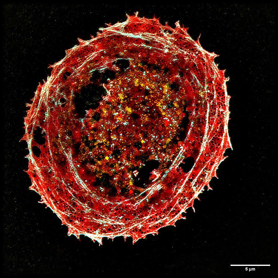
The novel optical setup provides an image of multicolor-stained liver cells. The image shows the cells’ tiny membrane structures, which are smaller than the diffraction limit of light. [Image: Henning Ortkrass, Bielefeld University] [Enlarge image]
Researchers in Germany and Norway have demonstrated a fluorescence microscope that achieves high-speed super-resolution imaging over a wide field of view (Opt. Express, doi: 10.1364/OE.495353). The instrument, which has been designed to deliver high-resolution images of cellular dynamics at video frame rates, will be used to probe the complex interactions between different drugs inside the body.
A broad (and rapid) view
The microscope is based on an established super-resolution imaging technique that exploits structured light patterns to excite fluorescence in a sample, ensuring that the excitation power remains low enough to avoid damaging living cells. The technique, called super-resolved structured illumination microscopy (SR-SIM), already delivers the spatial resolution and speed needed to study cellular dynamics, but existing instruments are bulky and offer only a limited field of view.
In contrast, the compact design developed in this new work can image areas of up to 150 × 150 µm2 with no loss of quality, while still achieving a resolution of less than 100 nm and a frame rate of 44 Hz. “With this new microscope, individual drug combinations can be tested on isolated cells and then imaged with super-resolution to observe the dynamics of cell membrane features or organelles,” said first author Henning Ortkrass, Bielefeld University, Germany. “The large field of view can provide statistical information about the cells’ response, which could be used to improve personalized health care.”
Studying cellular dynamics
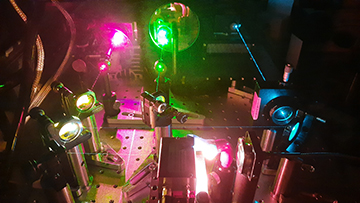
The super-resolution microscope exploits an innovative optical design to achieve high-speed imaging over a wide field of view. [Image: Henning Ortkrass, Bielefeld University]
The large field of view is achieved by using six optical fibers mounted on a custom-built hexagonal holder to illuminate the sample with the interference patterns needed for SR-SIM. The fiber holder collimates and refocuses the optical beams, while a fast-switching unit shifts and rotates its position to change the angle and phase of the light pattern.
The raw images acquired with this optical arrangement contain the high-frequency information needed to reconstruct high-quality super-resolved images, even when fluorescence is excited in multiple colors. The setup can also be tuned for total-internal-reflection fluorescence excitation, which achieves an even higher resolution by restricting the fluorescence excitation and detection to a thin region of the sample.
The researchers tested the microscope using cryopreserved samples of rat liver cells, with the reconstructed images revealing tiny membrane structures that are smaller than the diffraction limit of light. They now plan to use the instrument to investigate the dynamics of living cells after treatment with several drugs, and to improve the image reconstruction process to create super-resolved images from the raw data in real time.