
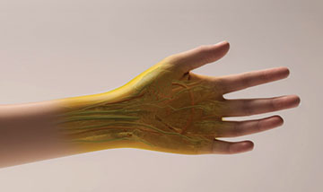
Researchers used food dye FD&C Yellow 5 to turn mouse skin transparent. They foresee this method being applied to humans in the near future. [Image: Keyi "Onyx" Li/US National Science Foundation]
The highly scattering nature of biological tissue poses an inherent challenge to the optical imaging of living animals. Current optical methods, such as two-photon microscopy and fluorescence imaging, are limited to superficial depths and low resolutions or are simply not suitable for live-animal imaging.
Now, researchers at Stanford University, USA, have taken a unique approach to this longstanding issue by rendering a mouse’s tissue transparent with the help of a common yellow food dye (Science, doi: 10.1126/science.adm6869). Counterintuitively, the addition of strongly absorbing molecules achieved optical transparency in a live mouse, allowing them to see internal organs with the naked eye.
“Our method can improve existing optical imaging modalities to probe deep features that [could not] be visualized before in live animals,” said study author Zihao Ou, now at the University of Texas at Dallas, USA. “Beyond that, we envision the potential clinical translation of this invention for early disease diagnosis and health monitoring in humans.”
The Kramers-Kronig relations
Light scattering in biological tissue originates from the refractive index mismatches among its different components. Water has a low refractive index, while other biomolecules like lipids and proteins have a high refractive index. Ou, who studies self-assembly and nonequilibrium behaviors in soft-matter systems, and his colleagues thought about applying dye to the skin to more closely match these refractive indices.
“Similar to the physics behind an oil–water mixture, light will be scattered in all different directions when propagating through the tissue, leading to optical opacity,” said Ou. “We leveraged one of the most well-known theories in optics, the Kramers–Kronig relations, to address this challenge.”
The Kramers–Kronig relations―equations that describe the relationship between absorption and refractive index―helped the researchers predict that the use of a dye that sharply absorbs in the near ultraviolet and blue regions would alter the refractive index at longer wavelengths when dissolved in water. According to their theoretical work, the introduction of strongly absorbing yellow dye molecules would effectively reduce the refractive index differences in biological tissue.
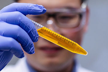
Zihao Ou holds a vial of the common yellow food coloring tartrazine in solution. [Image: The University of Texas at Dallas]
From mice to humans?
To test their hypothesis, the researchers rubbed a solution of red tartrazine, also known as the food dye FD&C Yellow 5, onto the abdomen, scalp and hindlimb of a live mouse. They were able to observe fluorescent protein–labeled enteric neurons in the gut, cerebral blood vessels and muscle sarcomeres, all with the naked eye. The reversible transparency also enabled them to view the mouse’s functioning internal organs, including the liver, small intestine, cecum and bladder.
Ou and his colleagues look forward to other researchers using their technique to enhance existing optical imaging methods. But they also foresee skin transparency being applied to humans in the near future, where it could replace or augment certain medical tests that are normally invasive or require radiation, such as X-rays and CT scans.
“With all the preliminary demonstrations, we hope to further improve the technique in multiple aspects, including dye synthesis, more efficient and accurate drug delivery strategy, and recipe design to achieve an optimal imaging condition with minimum adverse effects,” said Ou.