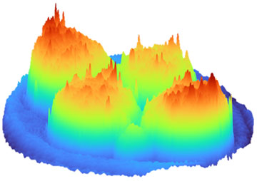
Mapping the refractive index of an embryo—in this case, at the four-cell stage of development—offers a fast and noninvasive way to assess its health as it develops, according to the latest study. [Image: University of Adelaide]
Researchers at the University of St. Andrews in the United Kingdom and the University of Adelaide in Australia say they have for the first time captured 3D holographic images of mouse embryos that provide extra information about their viability for in vitro fertilization, or IVF (Biomed. Opt. Express, doi: 10.1364/BOE.492292). By relating physical parameters extracted from the holographic data to a key indicator of embryo health, the technique could provide a rapid and noninvasive tool for identifying the best candidates for implantation.
Mapping embryos’ refractive index
While IVF clinics routinely examine the developing embryos to assess their health and growth potential, such visual inspections lack any quantifiable information to help guide the selection process. “Optical technologies hold immense promise to unravel the metabolism and health of the embryo,” says project coleader Kylie Dunning, University of Adelaide. “This gentle and noninvasive approach could lead to improved IVF success rates, which have remained stagnant for more than a decade.”
The researchers used a technique called digital holographic microscopy (DHM), which has become a popular method for studying the physical properties of living cells, to map the refractive index of mouse embryos at five different stages of development. Any change in the refractive index indicates that the embryo is accumulating more lipid-containing fluid, which is known to compromise the health of the developing organism.
Low power and label free
To test their approach, the researchers cultured mouse embryos in both low-lipid and high-lipid environments. The phase information contained in the digital holograms revealed dynamic changes in the refractive indices of the embryos, with the high-lipid group showing an elevated refractive index at almost all stages of development. In contrast, a normal visual examination showed no sign of any difference in lipid abundance between the two cohorts.
The researchers point out that DHM requires only very low excitation powers over short periods of time, which earlier studies suggest should not degrade the viability of the developing embryos.
The refractive indices of both groups dropped dramatically at the last development stage, which the researchers attribute to a fundamental change in the evolving embryo’s cellular structure. As an independent check, the team also confirmed that embryos cultured in the high-lipid medium had accumulated more fluid within their cells than those developed in a low-lipid environment.
The researchers point out that DHM requires only very low excitation powers over short periods of time, which earlier studies suggest should not degrade the viability of the developing embryos. The label-free nature of DHM also allows it to be used alongside other optical techniques, such as Raman spectroscopy and hyperspectral imaging, which could support a suite of noninvasive diagnostics to augment conventional visual assessments of embryo quality.
