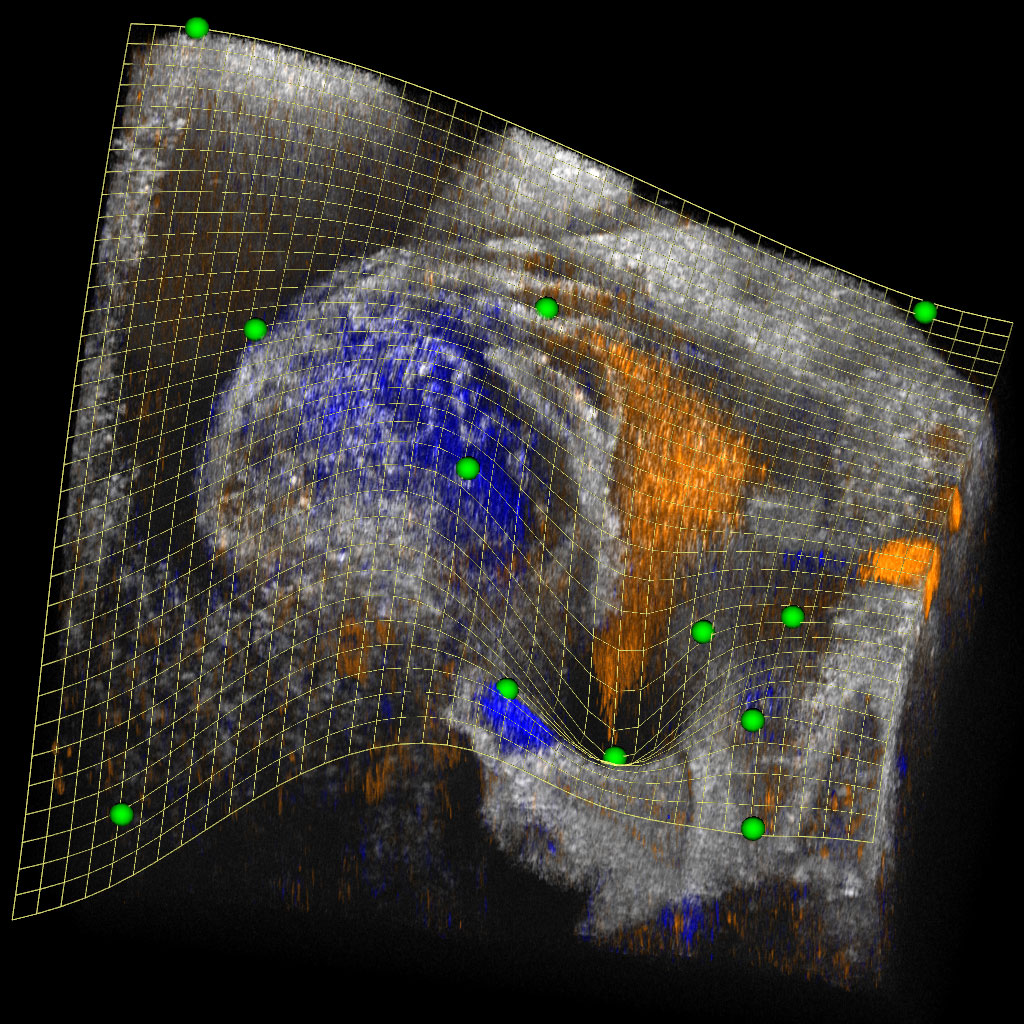
The clipping spline (mesh) is defined by the control points (green) and provides a smooth cutaway view into the looping embryonic mouse heart, revealing the blood flow (orange and blue) in the heart’s atrium and ventricle. [Image: Andre C. Faubert and Shang Wang, Stevens Institute of Technology]
Visualization of a 3D image allows it to be viewed on a screen, most often by displaying a 2D slice through the volume along a plane. However, planes are inherently limited in their capabilities, leading to sharp edges in the clipped volume and an inability to render concave surfaces.
Now, researchers at the Stevens Institute of Technology, USA, have created a new open-source software tool that offers improved 3D and 4D visualization of biomedical images (Biomed. Opt. Express, doi: 10.1364/BOE.544231). The tool, called the clipping spline, can be used with any optical imaging technique that produces volumetric images, such as optical coherence tomography (OCT), photoacoustic tomography, light-sheet microscopy, optical projection tomography and confocal microscopy.
Toward sophisticated visualization
When working with 4D OCT images of an embryonic mouse heart, Shang Wang and his colleagues had trouble visualizing the changes in cardiac morphology during development. Cardiac looping is a critical event that involves bending and twisting of the heart tube, going from straight to a more complex structure reminiscent of the adult heart. The researchers found only limited tools available for more sophisticated visualization, so they built their own.
“This specific need from our own research led to our creation of the clipping spline. Furthermore, similar needs widely exist from the biomedical imaging community,” said study author Wang. “With 3D and 4D images increasingly acquired and used, having a tool to visualize and analyze the targeted structures inside the volumes is simply critical for turning images into insights.”
In contrast to traditional clipping planes, the clipping spline produces non-planar cutaway views with an easily defined smooth surface that can follow a convoluted structure, thus fully revealing the structure in a single view. Also, in comparison to non-planar, voxel-centric methods, the clipping spline has the unique advantages of being fully adjustable and dynamic.
In contrast to traditional clipping planes, the clipping spline produces non-planar cutaway views with an easily defined smooth surface that can follow a convoluted structure, thus fully revealing the structure in a single view.
An open-source tool
The new tool leverages a type of 3D smooth surface known as the thin plate spline and applies it to volume clipping for the first time. A thin plate spline uses an unordered set of three or more control points to define the unique surface, which coincides with all control points while having minimal curvature. This surface is fully adjustable, and users can modify control points to refine its shape and position interactively.
“One can simply add, delete and move control points, and see the cutaway view in real time to optimize or change the visualization,” said Wang. “The cutaway viewing states can be automatically connected with smooth transitions, enabling 4D volume clipping and generating smooth flythroughs to reveal structures inside the volume.”
Wang and his colleagues demonstrated the clipping spline with 3D and 4D images from their own lab, revealing a series of never-before-seen dynamics and processes of embryonic mouse heart development at the looping stage based on OCT data. By making the tool publicly available and free to use, they hope to see other researchers incorporate the clipping spline into their workflows to improve visualization and advance biomedical research.


