
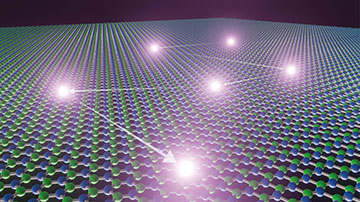
Researchers in Switzerland and the United Kingdom have shown how molecules in organic solvents can activate defects in sheets of boron nitride, causing the defects to emit single photons. This artist’s impression shows the trajectory of a solvent molecule as it hops from one defect to the next. [Image: Titouan Veuillet / EPFL]
Liquids confined to nanoscale spaces can experience remarkable changes to their structure and flow compared to bulk fluids. Imaging their behavior at such tiny scales is typically done by measuring the flux of molecules leaving the nanostructures in question rather than looking directly at what goes inside. The inability to count individual molecules means that scientists must average over large molecular ensembles, which leaves them reliant on theoretical models to describe flow and other fluid properties.
Now scientists in Europe have shown how to follow the dynamics of molecules in organic solvents by measuring the single photons emitted when the molecules interact with individual defects in hexagonal boron nitride (Nat. Mater., doi: 10.1038/s41563-023-01658-2). They have found that the spectrum of emitted light depends not only on the type of liquid present but also on how tightly confined that liquid is.
Light-emitting defects
Nanoscale matter can be probed optically via defects in solid-state materials, such as nitrogen vacancy centers in diamond, but such systems are difficult to embed in two-dimensional structures. Hexagonal boron nitride (hBN) is a promising alternative, as it consists of layers of graphene-like one-atom-thick sheets, with defects in the crystal structure that can act as room-temperature quantum emitters. Indeed, scientists have already shown how to activate these emitters by irradiating the material or doping it with carbon atoms.
In the latest work, Aleksandra Radenovic and Nathan Ronceray at the École Polytechnique Fédérale de Lausanne (EPFL), Switzerland, Boya Radha at the University of Manchester, UK, and colleagues have taken a different approach—immersing hBN in suitable liquids.
They found that while most emitters light up for just a few tenths or hundredths of a second, some remain active for several seconds.
The researchers peeled off flakes from high-purity crystals of hBN (using adhesive tape) so that the flakes fell on a glass coverslip that they placed in a chamber filled with one of several liquids. They then used a green laser to try and get the crystals to fluoresce. They were unable to do so with water, but when they switched to ethanol, the crystals generated an intense emission. The team observed similar results when using other common organic solvents, including pentane and methanol.
Attributing the emission to the solvents’ activation of defects already present in the crystals, the researchers then sought to document the activity of individual emitters. They did so by recording videos of a tiny portion of the crystal surface while blasting it with laser light, and then overlaying 5,000 of the captured frames. They found that while most emitters light up for just a few tenths or hundredths of a second, some remain active for several seconds. And in many cases, activated defects go on to stimulate neighboring defects, leading to wiggly patterns of fluorescence on the composite image.
Investigating the fluorescence
Radenovic and colleagues also looked at the emitters’ spectral response, using a technique known as spectral single-molecule localization microscopy. They found that the recorded spectra depended on the liquid used in each case, with more-polar solvents generating longer-wavelength outputs. This was true both for excited defects that dropped straight back down to their ground states and for those that did so after losing some of their energy to lattice vibrations.
The researchers admit that they don’t know exactly what is responsible for the emission, but they argue that defects are likely activated by charge being transferred to and from the solvent.
The researchers admit that they don’t know exactly what is responsible for the emission, but they argue that defects are likely activated by charge being transferred to and from the solvent. Neighboring defects then become activated as charge-bearing solvent molecules move across the crystal surface—with the resulting fluorescence pattern tracking the random walk of individual molecules through the liquid. The team points out that neither pristine hBN nor the solvents possess electronic transitions in the visible portion of the spectrum so deduces that defects become activated thanks to an “electronic structure rearrangement.”
To prove that the fluorescence they measured came from individual rather than groups of defects, the researchers recorded the light using two single-photon detectors. They found that the arrival times of individual photons were equally spaced from one another—displaying what is known as antibunching, a characteristic of single-photon emission. The researchers say that their optical readout therefore “truly reports on nanoscale properties of the liquid.”
Activation along nanoslits
To complete their work, Radenovic and colleagues switched from bulk liquid to its confined counterpart. They did so by creating two-dimensional rectangular “nanoslits” inside a three-layer structure: hBN on the bottom, a few sheets of graphene in the middle and a transparent layer of the mineral mica on top. They made each slit—which measured 150 nm wide and just one or two nanometers deep—by removing the graphene within it so that a molecule was confined by the hBN below, mica above and walls of graphene on the sides. Because graphene quenches light, only the slits yielded emission when a liquid passed through the structure.
As with bulk liquid, the researchers showed that they could activate a chain of emission along the nanoslits.
As with bulk liquid, the researchers showed that they could activate a chain of emission along the nanoslits. But they also found that the wavelength of that emission depended on precisely how deep the slits were. Reducing the height of the slit from 2.4 nm to 1.4 nm yielded a clear blueshift of the emitted radiation—changing the spectral signature of a strongly polar solvent so that it came to resemble that of a nonpolar alkane and implying that the dielectric constant drops in shallower channels.
The researchers point out that the single-molecule microscopy technique that they used is widely available and the organic solvents are commonplace, arguing that their new scheme could be applied to optical imaging and sensing of molecules, actively driven in nanoslits by electric fields or pressure differences.
Ronceray says that the approach is most likely to aid fundamental research, but he reckons it could also help shed light on how liquids behave in 2D material–based membranes—which might in future be used in desalination plants. He adds that he doesn’t foresee any particular technical barriers to the scheme’s adoption but rather a sociological one—a lack of overlap between the microscopy and nanofluidics communities. “Labs may need to team up,” he says.