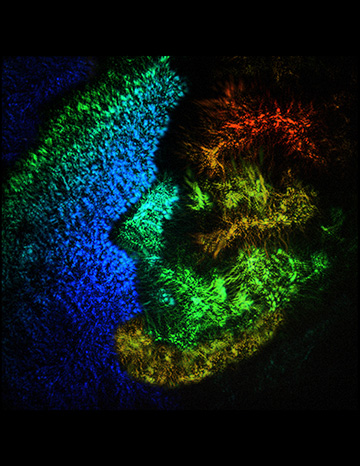
The portrait of Lincoln on the face of a U.S. penny is visualized using a new tomographic mid-infrared imaging technique from the University of California, Irvine, USA. Depth resolution is enabled by adjusting the time delay between the mid-IR and gate pulses at the camera chip, while axial resolution is determined by the short coherence length of the femtosecond pulses. The 3D data stack can, according to the authors, be effectively measured just under 1 second. [Image: Fishman/Potma/Knez]
Tomographic imaging with mid-infrared (mid-IR) light has emerged in recent years as a way to visualize the chemical composition of samples without the need for molecular contrast agents. Most molecules of interest display fundamental vibrational absorptions in the mid-IR, which allows for their identification through distinctive spectral fingerprints.
However, progress in this field has been hindered by the lack of fast, low-noise and cost-efficient mid-IR cameras. Researchers from the University of California, Irvine (UCI), USA, have overcome this technical hurdle by developing an approach to mid-IR tomography that uses a conventional silicon-based CCD camera (Optica, doi: 10.1364/OPTICA.426199). The technique enables wide-field, high-definition 3D imaging with acquisition rates up to two orders of magnitude higher than seen with previous methods.
An ordinary CCD camera
Mid-IR detector technology has remained fairly stagnant, with fast cameras having a very small number of pixels and with construction often based on low-band-gap materials that suffer from high thermal noise. To advance mid-IR tomography, scientists needed to find alternatives to such limiting technology.
“Because of this, there is an idea—and this is not a unique idea—about how we can convert information from the infrared range to the visible range,” said Dmitry Fishman, director of laser spectroscopy labs in the UCI department of chemistry. “There is an orchestrated group effort to do this using different approaches, including converting this information on the sample. What we offer is a different approach: the idea of conversion of the information right on the camera chip.”
Fishman and his colleagues realized they could use the process of nondegenerate two-photon absorption to achieve mid-IR detection with an ordinary, off-the-shelf CCD camera. Essentially, a mid-IR pulsed beam passes onto a sample, and the reflected or scattered light is collected by a lens that projects an image of the sample onto the CCD. When this image is overlapped with a separate near-infrared pulsed beam, a voltage is detected by the CCD.
New possibilities for mid-IR tomography
The researchers went a step further by using the pulse overlap as a temporal gate to achieve depth resolution. The gate only allows for the detection of light that has traveled a preset path length, corresponding to a certain depth of the sample. The team successfully obtained 3D images of a coin through both transparent and highly absorbing media. Also obtained were images of polymer structures and protein crystals with spectroscopic contrast to showcase chemical selectivity.
“The main advance that we put on the table is that you can take a regular CCD camera and, number one, turn that camera into one can now see things in the mid-IR,” said Eric Potma, a professor of chemistry at UCI. “Now, you can use that camera not just for 2D images, but 3D images of an object in the mid-infrared. That of course is very important because the mid-infrared has this extra information about chemical composition.”
The researcher believe that the boost in speed and simplicity opens up new possibilities for mid-IR tomography, particular in the widespread application of non-destructive and non-contact materials inspection. For instance, electronics manufacturers could visualize the insides of devices without having to open them up, or makers of aerospace components could spot any structural weaknesses that might lie under the surface.
