
 [Getty Images]
Understanding the brain has been a grand challenge for decades. Although medical imaging technology (such as magnetic resonance imaging) has allowed scientists to image the brain on a macroscopic level, there is still much to be learned at a finer resolution. Optical microscopy can provide this resolution, allowing the imaging of individual neurons. Fluorescence-based optical imaging even enables researchers to visualize functional dynamics—for example, to see if individual neurons are firing or not—by using functional indicators that change brightness with neuronal activity.
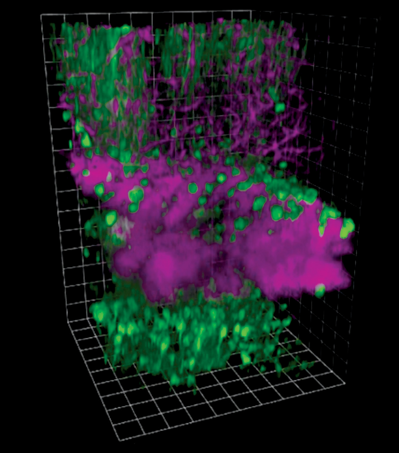 [Enlarge image]3D reconstruction of three-photon images of GCaMP6s-labeled neurons in the mouse cortex and hippocampus. Fluorescence (green) visualizes neuronal bodies and temporal neuronal dynamics, while third-harmonic generation (magenta) visualizes blood vessels and myelinated axons. [Adapted from Nat. Methods 14, 388 (2017)]
Despite the increased resolution, traditional one-photon microscopy only allows for a fraction of the brain of even small laboratory mice to be imaged—it can only image neurons up to a certain depth. The obvious goal is to extend the depth at which fluorescence-based optical microscopy can operate, and multiphoton microscopy has become a popular solution.
Beyond one photon
Multiphoton microscopy works similarly to other fluorescence microcopy–based techniques. The cells of interest are labeled with fluorescent molecules, and a scanning laser beam is used to excite these fluorophores. In multiphoton microscopy, this excitation is performed using more than one photon. In n-photon excitation, n photons (each with ~1/n the photon energy of the photon energy used for one-photon excitation) combine simultaneously to excite the fluorophore. Because this process is nonlinear and much weaker than one-photon excitation, multiphoton excitation requires the use of an ultrafast laser to enhance the excitation probability while maintaining moderate average power levels. Although using an ultrafast laser is cumbersome, multiphoton excitation provides major advantages in deep tissue fluorescence microscopy.
The highly scattering nature of biological tissue means that the ballistic excitation photons have a reduced probability of reaching the focus and exciting the fluorophores. The attenuation of ballistic photons in a scattering medium can be quantified by a scattering coefficient, which is typically smaller for longer wavelengths. Because multiple photons combine their energy to generate one molecular excitation, multiphoton microscopy uses a much longer excitation wavelength than one-photon excitation of the same fluorophores. For example, performing two-photon microscopy on visible-light-emitting fluorophores requires near-infrared excitation wavelengths, which allows better delivery of the excitation photons over a larger length and thus improves ballistic photon penetration in tissue.
Another advantage is that the nonlinear nature of multiphoton excitation provides intrinsic excitation confinement in all three dimensions—most of the fluorescence is generated within a small volume around the focus. Combined with laser scanning, this excitation confinement allows unambiguous assignment of the generated fluorescence to its origin, even if the emitted fluorescence is scattered by the tissue.
The obvious goal is to extend the depth at which fluorescence-based optical microscopy can operate, and multiphoton microscopy has become a popular solution.
Researchers extended imaging depth by turning to long-wavelength two-photon fluorescence microscopy, using excitation wavelengths greater than 1.2 µm, where the scattering coefficient is smaller. However, introducing longer wavelengths presents its own challenges. First, the high water content of in vivo tissue means that absorptive loss becomes important for wavelengths greater than 1.2 µm, and one must consider the tradeoff between scattering (coefficient αs) and absorption (coefficient αa). This is done by defining an effective attenuation coefficient, αe = αs + αa, which has its smallest value at ~1.7 µm and local minima at ~1.3 µm and ~2.2 µm for in vivo mouse brain tissue. This shows that the ideal excitation wavelength for the mouse brain is ~1.7 µm.
The second consideration is that this wavelength is too long for nearly all fluorophores to be excited with two-photon microscopy. Fortunately, ~1.7 µm is compatible with three-photon excitation of red fluorophores, and the local minimum at ~1.3 µm is compatible with three-photon excitation of the most biologically important green fluorophores. The ~2.2 µm local minimum is impractical unless four-photon excitation is used.
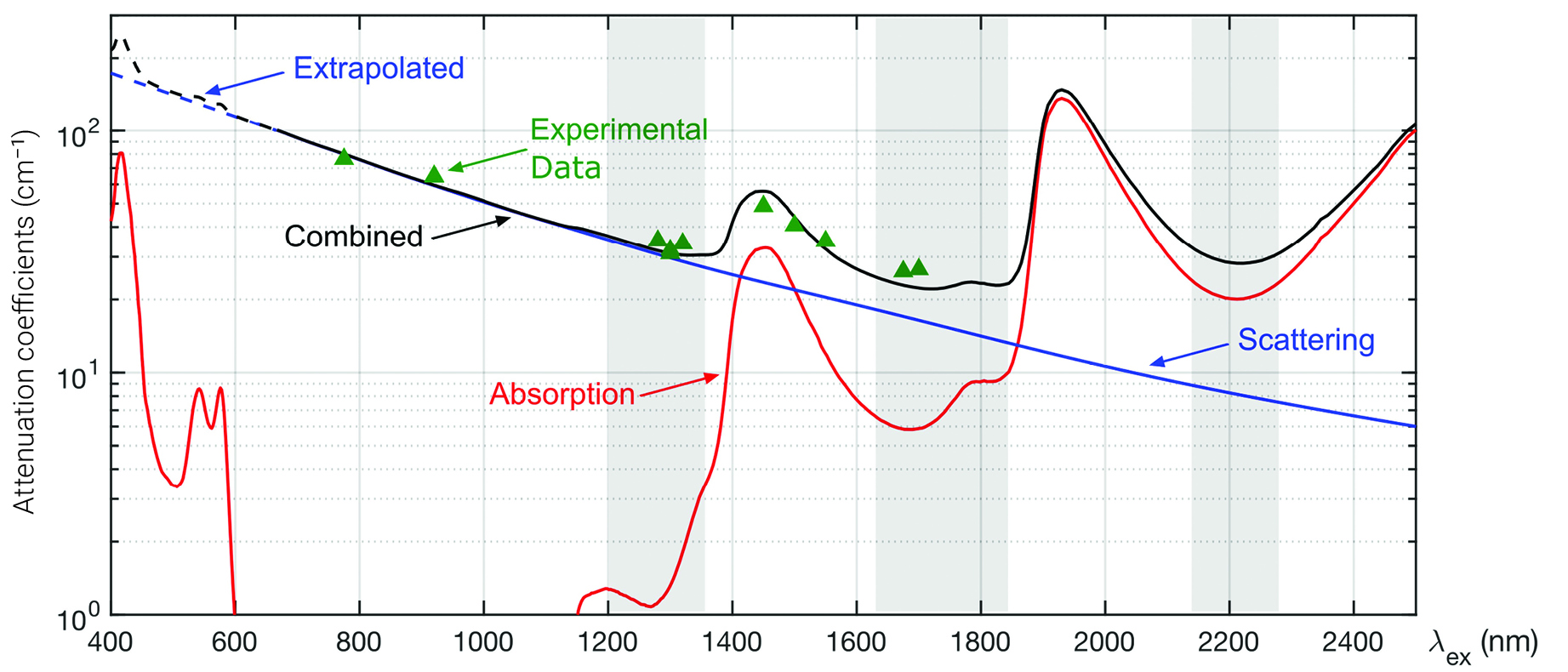 [Enlarge image]Absorption coefficient (red), scattering coefficient (blue) and effective attenuation coefficient (black) of the in vivo mouse brain plotted as a function of wavelength. Dashed lines indicate where data have been extrapolated. Green triangles are experimental measurements of the mouse brain. Grey highlighted regions show the long-wavelength windows for deep tissue imaging. [Adapted from J. Phys. Photonics 4 (042501), 1 (2022)]
Three-dimensional excitation confinement is also essential for deep tissue imaging. Although scattered excitation photons do not contribute to fluorescence at the focus, they can excite fluorophores elsewhere, producing background fluorescence that ultimately leads to loss of contrast. A quantitative measure of the image contrast is the ratio of the in-focus signal to out-of-focus background, referred to as the signal-to-background ratio (SBR).
Although the nonlinear nature of multiphoton excitation confines the excitation to the focus (neglecting the presence of scattering), theoretical analysis and experimental studies have both shown that the SBR eventually approaches zero when imaging deep in scattering tissue. Because of its higher-order nonlinear excitation, the SBR of three-photon microscopy is much greater than that of two-photon microscopy, but at the expense of signal strength due to the higher order of the nonlinear process. Currently, the signal strength rather than SBR limits the depth of three-photon imaging.
The increased nonlinearity of three-photon excitation also means it requires a higher pulse energy than two-photon microscopy, and a lower-repetition-rate laser must be used to stay below the safe average power level for biological tissues. Consequently, standard three-photon microscopes generally operate at a lower frame rate than two-photon microscopes (since at least one pulse must be present per pixel).
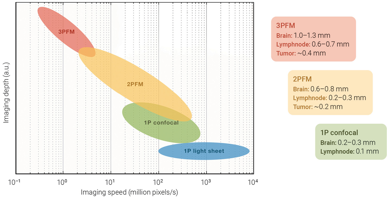 [Enlarge image]Left: Tradeoff between imaging depth (arbitrary units) and imaging speed for three-photon fluorescence microscopy (3PFM), two-photon fluorescence microscopy (2PFM), one-photon (1P) confocal and 1P light sheet. Right: Examples of biological tissues that have been imaged with each technique and the depth for typical green fluorophores. [Adapted from Cell 187, 4458 (2024)]
The search for a source
The biggest challenge to making three-photon imaging a robust, practical tool for deep tissue imaging has been the development of a reliable excitation source that meets the need for a higher pulse energy and lower repetition rate as previously described. When considering losses in the microscope, a practical source would need to produce ultrafast pulses with ~1 µJ pulse energy at ~1 MHz repetition rate.
The first viable source, developed about a decade ago, was based on soliton self-frequency shift in a photonic crystal rod pumped with a commercial erbium-doped fiber laser. It was able to produce MW peak power pulses (~70 nJ pulses at ~70 fs pulse width) at ~1.7 µm with a 1 MHz repetition rate, allowing for the first deep three-photon images to be taken of neurons labeled with red fluorophores.
Despite this success, the wavelength of this laser is not compatible with three-photon excitation of green fluorophores, so scientists pushed to develop a source at ~1.3 µm. An early approach used a high-repetition-rate Ti:Sapphire regenerative amplifier to pump an optical parametric amplifier (OPA), producing ~400 nJ pulses at 1.3 µm. But this bulky system’s low repetition rate of 250 kHz, combined with the higher loss of external dispersion compensation optics and larger αeat 1.3 µm, meant it barely enabled recording neuronal activity beyond the reach of a two-photon microscope.
A similar approach instead used a frequency doubled Yb (ytterbium)-doped fiber master oscillator power amplifier (MOPA) to pump a noncollinear OPA (NOPA). This method provided 1.25 µJ pulses at 1.3 µm with a repetition rate of 400 kHz (which could be externally doubled to 800 kHz) and ultimately allowed recording of high-fidelity neuronal activity traces in the mouse hippocampus without removing the superficial regions of the brain. Starting around 2016, commercial NOPA systems have been improved upon by laser companies including Coherent, Spectra-Physics, Light Conversion, Class 5 and others, and these companies are now selling systems that can provide more than 1 µJ pulse energy at greater than 1 MHz repetition rates at 1.3 µm. Since the NOPA is wavelength tunable, the Yb pumped NOPA has become the standard system for imaging at ~1.7 µm as well.
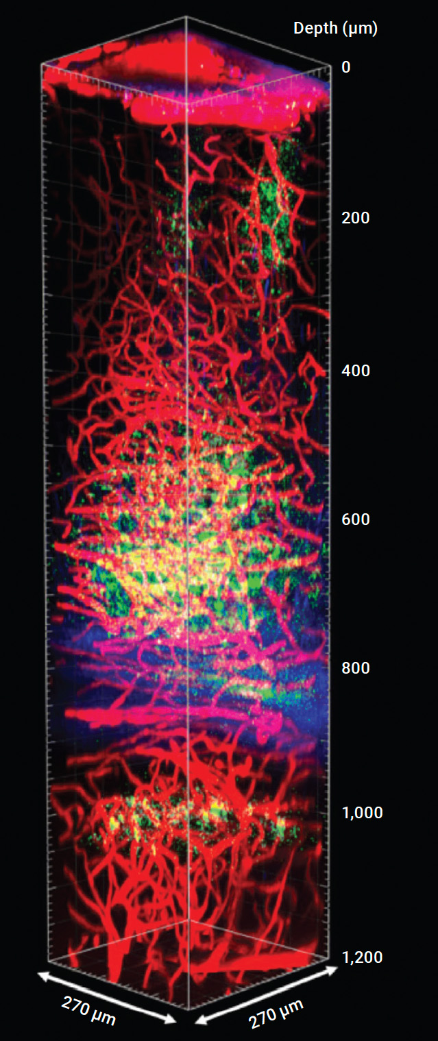 [Enlarge image]Multicolor three-photon Z-stack image down to 1.2 mm below the surface of the brain, showing Texas Red–labeled blood vessels (red), GCaMP6s-labeled neurons (green) and third-harmonic generation (blue). [Adapted from Sci. Adv. 7, eabf3531 (2021)]
Advances in imaging
Given that three-photon microscopy calls for either a ~1.3 µm or ~1.7 µm excitation source, generating multicolor three-photon images would appear to require using multiple excitation wavelengths (for example, ~1.3 µm for green and ~1.7 µm for red fluorophores). Although the multiple-wavelength approach has been demonstrated, it is more complicated than using a single excitation wavelength. Recently, however, researchers demonstrated multicolor single-wavelength excitation at 1.34 µm.
The key mechanism this approach exploited was three-photon higher-energy excited state excitation of red fluorophores. In this scheme, the energy of two of the three photons nearly bridges the energy gap between the ground and lowest excited state. The third photon bridges the gap and subsequently excites the fluorophore, but to a higher-energy excited state. Due to the higher photon energy in this scheme, shorter excitation wavelengths can be used, enabling simultaneous “conventional” three-photon excitation of green fluorophores and “high-energy” three-photon excitation of red fluorophores.
Quantum perturbation theory also predicts that higher-energy excited state excitation may cause resonance enhancement, and the three-photon cross-section may be higher (as compared with conventional excitation to the lowest excited state). Measurement of common red fluorophores has shown that enhanced blue-shifted cross-sections exist for several of them, indicating that they may be undergoing resonance enhancement. Recent measurements of fluorescein, a green fluorophore, have also shown enhanced blue-shifted cross-sections, indicating that green fluorophores may also show resonance enhancement. Practically, these enhanced cross-sections make the single-wavelength multicolor scheme even more attractive. They can sometimes be more than 10 times the three-photon cross-section at the energy scaled one-photon excitation peak. This development may prove useful in extending the depth of three-photon imaging, as it is currently limited by the signal strength.
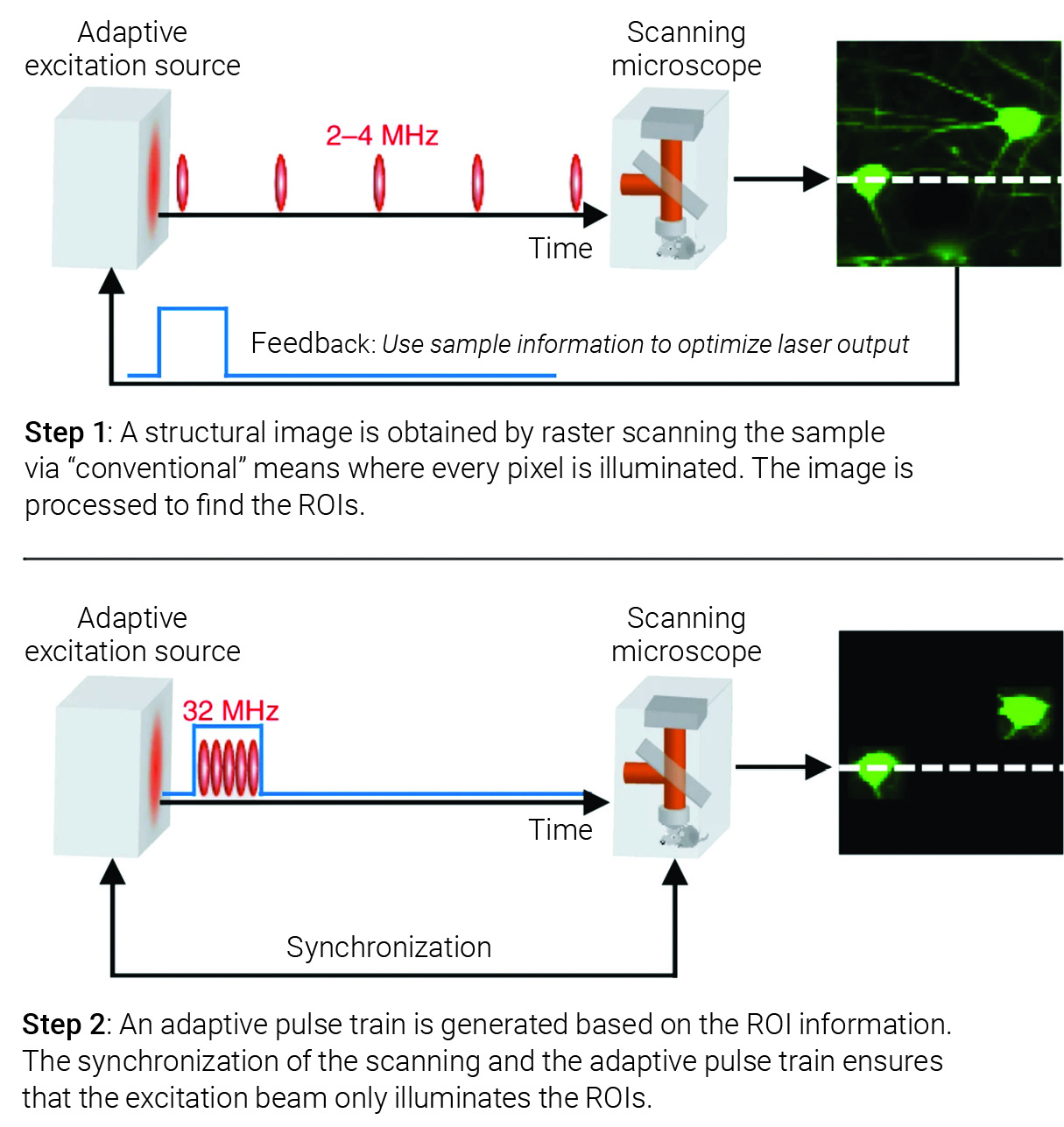
Working principle of a pulse-on-demand system (for example, the adaptive excitation source). [Adapted from Nat. Methods 17, 163 (2020)]
One approach to increasing the signal strength is using “pulse-on-demand systems” such as the adaptive excitation source. The idea of adaptive excitation is simple: only illuminate the places in the field of view that contain the regions of interest (ROIs). This allows for a low average power over the entire field of view, but the pixels within the ROIs can be illuminated with higher pulse energy or additional pulses, improving the signal strength obtainable compared with non-adaptive excitation for the same volume rate (the rate at which the entire volumetric field of view is imaged).
Forming a sharp focus is essential for deep tissue imaging, so several groups have explored the use of adaptive optics to increase signal strength and spatial resolution. The idea is that since biological tissue aberrates the excitation point spread function (PSF), and n-photon excitation is proportional to the intensity raised to the nthpower, then if the optical aberrations can be corrected, a higher peak intensity will be present at the focus, increasing the generated signal.
In addition, reducing the side lobes of the PSF caused by tissue aberration can improve spatial resolution and SBR. Since methods for direct measurement of aberrations only work for transparent or moderately scattering tissues, indirect and computational approaches for correcting the PSF have been developed. So far, these techniques have demonstrated a roughly 10× increase in signal for three-photon imaging of neurons, and even greater gains for smaller structures—for instance, about 30× for dendritic structures.
Given these advances, researchers can now image mouse brain vasculature in vivo at a depth greater than 2 mm with long-wavelength two- and three-photon microscopy, and have imaged neuronal activity in the mouse brain at a depth greater than 1 mm and at a time resolution of ~10 Hz. There have also been advances in imaging the in vivo brain in a more “natural” way. For example, imaging can be done through the intact mouse or adult zebrafish skull with single-cell spatial resolution, ~10 Hz time resolution, and a field of view (FOV) of hundreds of micrometers, which removes the need to perform an invasive craniotomy. Additionally, head mounted two- and three-photon “miniscopes” allow for deep tissue recording of neuronal activity in freely moving rodents. In a push to see more neurons, deep tissue imaging has been combined with techniques such as large-FOV imaging and high-speed imaging to record the activity of a large number of neurons.
Forming a sharp focus is essential for deep tissue imaging, so several groups have explored the use of adaptive optics to increase signal strength and spatial resolution.
Although many multiphoton technology developments have focused on neurons in the brain, other non-neuronal cells, like astrocytes, microglial cells, oligodendrocytes and pericytes, or brain vasculature can be imaged and studied as well. Deep imaging technology has also extended beyond neuroscience to immunology and cancer research, including assessment for use in resecting tumors.
The ultimate depth limit
Multiphoton microscopy is fundamentally limited in depth by the out-of-focus background fluorescence. This background, in comparison with the in-focus signal fluorescence, causes a reduction of contrast in the image. When the SBR approaches zero, a usable image cannot be practically formed. Numerical and experimental work have shown that much of the background arises due to scattering, so as the imaging depth increases, one suffers not only from loss of signal due to the attenuation of ballistic photons, but also an increase (relative to the signal) in background photons and therefore a reduction of SBR.
The depth limit is generally defined as the depth where the SBR becomes unity. Although imaging beyond this limit is possible (for example, by integrating for a long time), it requires a prohibitive increase in the number of photons needed to generate an image with sufficient quality. The amount of background generated depends on both the effective attenuation coefficient and the labeling density (the density of fluorophores in the sample) and varies from tissue to tissue. In the mouse brain, the depth where SBR = 1 has been reached with two-photon imaging, but not with three-photon imaging. In three-photon in vivo imaging, the depth at which one can image is currently limited by the signal strength, suggesting that one could image deeper if a more intense signal were generated.
Looking ahead
One way to improve the practicality of deep three-photon imaging is to develop new excitation sources. Although the NOPA systems are practical, they are less competitive in cost and robustness than a typical laser source for two-photon imaging, mainly because of the conversion efficiency. Commercial NOPA systems can convert less than 5% of the laser power from the MOPA to ~1.3 µm, and usually less for conversion to ~1.7 µm (as a comparison, in a Ti:Sapphire laser, the conversion from the CW pump to the pulsed output is ~ 20% at the peak). Furthermore, as more pulse energy is required for deeper imaging, Yb MOPAs with higher pulse energy are required when imaging deeper.
Such a high-power pump not only increases the cost of the system, but also reduces robustness. And since there is a limit to the maximum average power that can be used for imaging, deeper imaging also requires a lower repetition rate. If a laser could provide constant average output power while varying the repetition rate, then the need for increased pulse energy with greater depth would not require a higher pump power. However, no laser currently has this capability, so the MOPA/NOPA can only be optimized for a specific imaging depth (either having pulses with more energy than necessary at shallow depths or not enough for deep imaging). There may be approaches that can improve conversion efficiency, such as compressing pulses before wavelength conversion or using gas-based nonlinear optics in hollow-core fiber.
Pulse-on-demand systems, such as the adaptive excitation source (AES), may also prove critical for reaching the depth limit where SBR = 1 in three-photon microscopy. These can either be realized by building a laser system that only produces pulses when needed to excite the sample, or by pulse picking. The first approach is more energy efficient but harder to realize (especially for arbitrary temporal patterns) than the second, again proving the need for more efficient laser sources.
Another tactic is to optimize the fluorophores themselves, increasing the signal strength by choosing fluorophores that have large three-photon cross-sections. Both red and green fluorophores have been shown to have blue-shifted resonance enhancements, and thus increases in their cross-sections, although only a handful have been rigorously examined. These experiments also revealed that the spectral shape of the three-photon cross-section cannot necessarily be predicted from energy scaling the one-photon cross-section. This indicates the need for a library of high-quality reference standards for both understanding the photo-physics and optimizing three-photon imaging. Combining enhanced cross-sections with adaptive optics and AES may also prove critical.
Other potential applications include imaging dense organs in vivo. Research would need to be done to identify how useful three-photon imaging would be in looking at organs with high blood content, like the liver, kidney or spleen, since blood is more scattering than the brain. Computational techniques such as artificial intelligence and denoising algorithms could also help push the limits of imaging depth, but here, too, a rigorous assessment is needed to determine how useful they are and where they may be most impactful in the quest to understand biological systems.
The size of biological tissue and the demand to see cellular functions in their native environment will drive the need for deep tissue imaging at high spatial and temporal resolution.
Deep tissue microscopy is a monumental task in optical imaging. During the last several decades, multiphoton microscopy, together with the accompanying development of lasers, microscope optics, detectors and fluorescent probes, has revolutionized our ability to image deep in scattering tissue. The size of tissue and organs and the demand to see cellular functions in their native environment will continue to drive the need for deep tissue imaging at high spatial and temporal resolution for the next decade and beyond.
This material is based upon work supported by the Under Secretary of Defense for Research and Engineering under Air Force Contract No. FA8702-15-D-0001. Anyopinions, findings, conclusions or recommendations expressed in this material are those of the authors and do not necessarily reflect the views of the Under Secretary of Defense for Research and Engineering.
Aaron K. LaViolette is with the Massachusetts Institute of Technology Lincoln Laboratory, USA. Chris Xu is with the School of Applied and Engineering Physics, Cornell University, USA.
For references and resources, visit: optica-opn.org/link/2504-multiphoton.
