
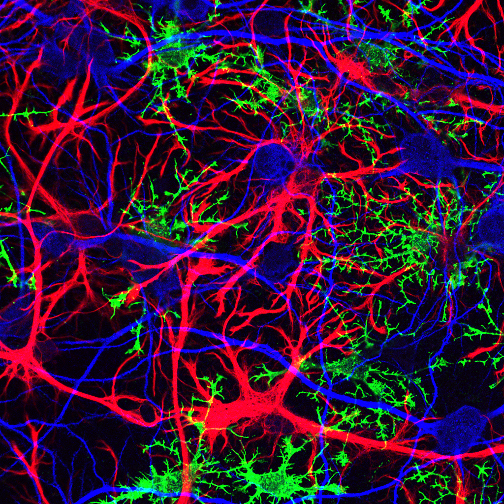
Rat hippocampus
Confocal microscopy of the rat hippocampus in culture: shown are immature oligodendrocytes (green, anti-NG2 and Alexa Fluor 488), astrocytes (red, anti-GFAP and Alexa Fluor 568), and neurons (blue, anti-MAP2 and Alexa Fluor 633) after 12 days in vitro. Image acquired on a Zeiss LSM 510 NLO with 40x 1.3 N.A. oil immersion lens.
—Jonathan Cohen, National Institute of Health, Bethesda, MD, USA