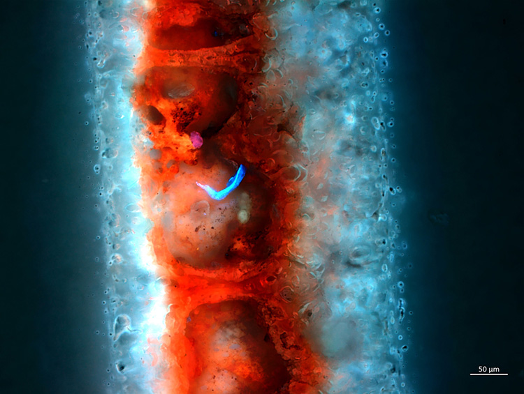
FIRST PLACE
“Fluorescence of a laser engraving mark on a white plastic card shows how the laser modified the molecules to create a contrast in the zones of the laser mark. Image details: 20x/0.5 objective, 12V 100W Halogen Lamp and filter set Ex400-440 BS460 Em 470-900. The 697×526-μm image is an extended focus from 45 Z-Slides (65.13 μm in total).”
—Jose Manuel Martinez Lopez, Quimica Tech, Chihuahua, Mexico
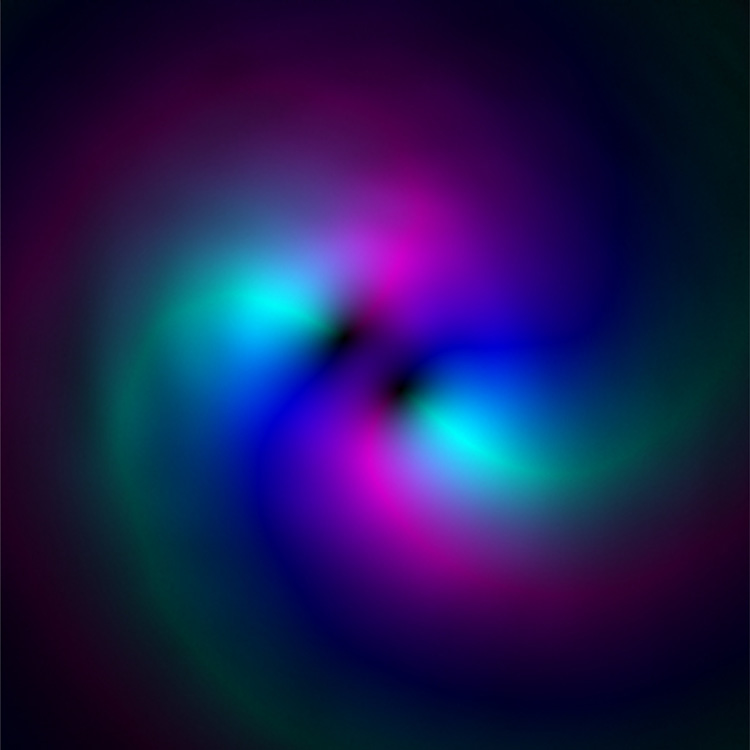
SECOND PLACE
“An orbital angular momentum (OAM) beam with charge L=2, reconstructed using the extended Gerchberg Saxton phase retrieval technique. In the image, brightness corresponds to intensity and hue to phase.”
—Matthew N. Jacobs, The University of Colorado Boulder, CO, USA
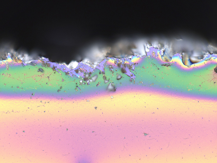
THIRD PLACE (Tie)
“Vivid snow summit with a blizzard in dark night sky. Actually, it is the irregular border of a graphene oxide (GO) film (~140 nm) spin-coated on a silicon substrate (300-nm SiO2/Si) and captured by the KEYENCE VK-X1000 Laser Microscope.”
—Zhijun Xu, College of Science, Zhejiang University of Technology, Zhejiang, China
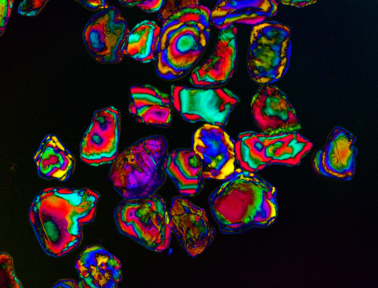
THIRD PLACE (Tie)
“White sand from Chapoquoit Beach (Falmouth, MA, USA) in transmitted light under polychromatic polarization microscope, 4× objective lens (nature.com/articles/srep17340).”
—Michael Shribak, Marine Biological Laboratory, University of Chicago, IL, USA

HONORABLE MENTION
“A snifter and a splash of cognac make a splendid liquid lens that captures summer solstice boating on the Foster City, CA, USA, lagoon with warmth and fascination. Spectral filtering was the intended purpose, but the variable Lagrange invariant across the field with the use of a Nikon D7100 now seems obvious in retrospect.”
—Dennis M. Hancock, Woodside, CA, USA
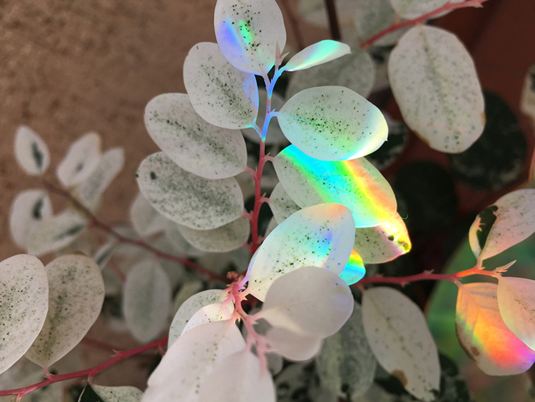
HONORABLE MENTION
“Sunlight diffracted and reflected by CDs over the white leaves of a Breynia distichia plant. Image taken with an iPhone 6s Plus without retouching.”
—Natalith Palacios-Ortega, Centro de Investigaciones en Optica, Mexico
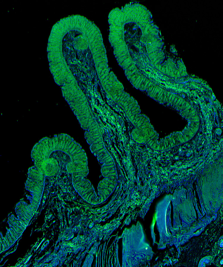
HONORABLE MENTION
“Composite image of a pre-cancerous colon section acquired on an infrared (IR) microscope reveals intricate structures that highlight underlying chemical composition. IR imaging enables label-free disease diagnostics using the intrinsic molecular contrast. The image was taken in the Chemical Imaging and Structures Laboratory headed by Prof. Rohit Bhargava at the Beckman Institute.”
—Yamuna Phal, University of Illinois at Urbana-Champaign (UIUC), IL, USA
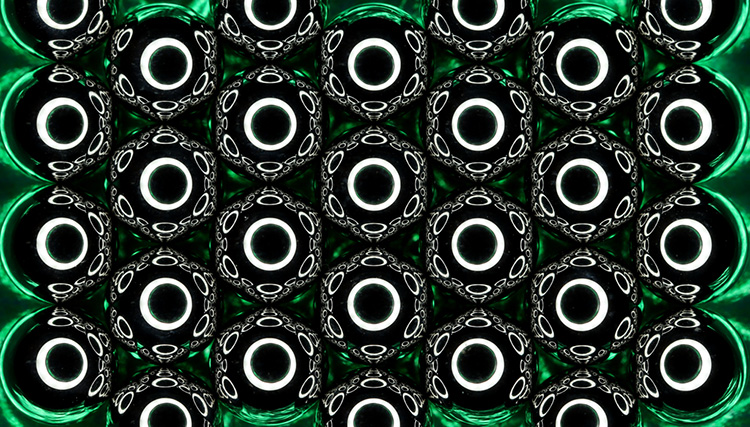
HONORABLE MENTION
“An array of 5-mm-diameter chrome steel ball bearings on a green foil is illuminated by, and photographed through, a single LED ring light placed a few centimeters above the array. Each ball bearing (acting as a convex mirror) forms a demagnified, virtual image of the 70-mm diameter ring light. This image is re-imaged by all the neighbouring ball bearings that can “see” it, and that image is re-imaged again, and again, ... ”
—Aongus McCarthy, Heriot-Watt University, Edinburgh, Scotland
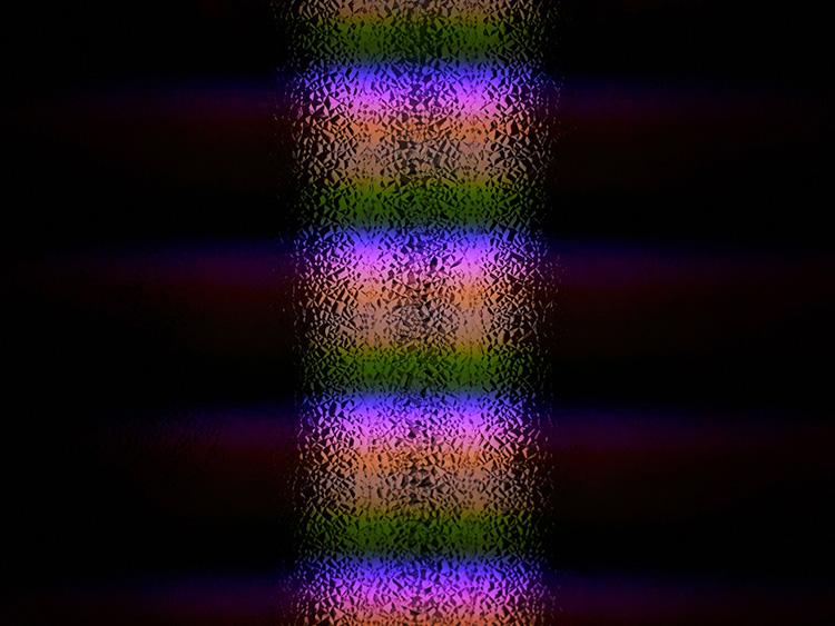
EDITORS' CHOICE
“Fluorescent tubes go through one on-off-on cycle every 1/120 second. Each row of pixels is exposed sequentially, adding up to 1/30 second or four full cycles going from top to bottom. Purple light emitted early in the cycle comes from mercury and short-lived states in the phosphor mixture, and green light emitted late in the cycle comes from a long-lived Tb(3+) state in the phosphor mixture that continues emitting light at 542 nm for several ms after mercury emission turns off.”
—Dan Deptuck, CMC Microsystems, Ontario, Canada
For this year’s contest, OPN received 54 remarkable entries. We thank the panel of judges who provided insight on those images and helped select the winners: Kate Bechtel, Felipe Beltrán-Mejía, Jennifer Kruschwitz, Anne Matsuura and Joel Villatoro.
You can see all of this year’s contest entries online at www.optica-opn.org/contest/2021.

