Researchers developed a "Molecular lantern" able to detect brain tissue altered by cancer or traumatic injury. [Image: PALMHELP/ Getty Images]
According to Greek lore, the ancient philosopher Diogenes carried a lantern to look for an honest man. Researchers in Spain, Italy and France are now using a tiny “molecular lantern” to seek changes inside the brain.
The European scientific consortium has developed a tapered optical-fiber probe that performs vibrational photometry inside the brains of mice to distinguish normal tissue from tumor metastases and traumatic brain injury (Nat. Methods, doi: 10.1038/s41592-024-02557-3). A deep-learning algorithm helps the spectroscopic system suppress background signals from the probe.
Brain probing: the optical paradigm
Over the last 20 years, scientists around the world have investigated various optical methods for probing the structure and health of living brain tissue. Although optogenetic techniques with embedded genetic tags can record neural activities, biochemical studies have lagged because the relevant molecules have broad emission spectra. Also, deep inside the brain, good signal-to-noise ratios are hard to come by.
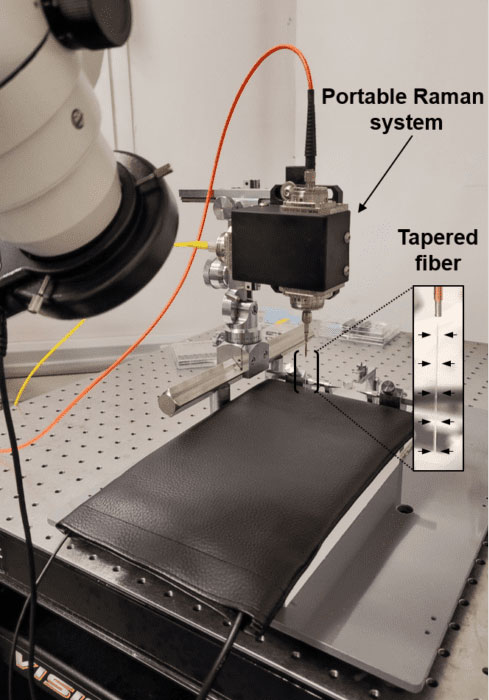
The vibrational spectroscopy instrument used in the “molecular lantern” experiments. [Image: Mariam Al-Masmudi, CNIO]
To overcome these hurdles, researchers have turned to techniques such as vibrational spectroscopy and Raman spectroscopy. These methods employ probes that are too small for the human brain—but their size is just right for the brains of mice, which are common neuroscience models.
A group from the NanoBright consortium, led by Filippo Pisano and Ferruccio Pisanello of the Istituto Italiano di Tecnologica, Italy, designed two label-free vibrational fiber spectroscopy tools to overcome the limitations of previous experiments. One was a bench-top system that provided the highest possible spectral resolution; the other was designed to be portable, for use in neuroscience laboratories.
“This technology allows us to study the brain in its natural state; it is not necessary to alter it beforehand,” said Manuel Valiente, National Cancer Research Center, Spain. “But it also makes it possible to analyze any type of brain structure, not only those that have been genetically marked or altered, as was the case with the technologies used until now. With vibrational spectroscopy we can see any molecular change in the brain when there is a pathology.”
Design and testing
After numerical simulations of possible interactions between the tapered optical-fiber probes and murine brain matter, the researchers designed tapered optical-fiber probes with a maximum diameter of 225 μm and a tip just 1 μm wide. For the bench-top system, the scientists attached the fiber probe to a custom confocal Raman microscope and a 785-nm-wavelength laser. A dichroic mirror performed the dual function of steering the laser light and sending the collected Raman signal to a spectrometer for analysis. For the portable system intended to examine living mice, the team placed three fiber ports on a single breadboard measuring 51 × 7.6 mm.
The group successfully used the technique to look at the epileptogenic zones surrounding traumatic brain injuries. “We were able to identify different vibrational profiles in the same brain regions susceptible to epileptic seizures, depending on their association with a tumor or trauma,” Liset M. de la Prida, Neuronal Circuits Laboratory, Cajal Institute, Spain, Prida explained. “This suggests that the molecular shadows of these areas are affected differently, and can be used to separate different pathological entities by automatic classification algorithms including artificial intelligence.”
According to the researchers, the spectroscopic systems are not yet ready for use on humans but hold great promise for studying animal models of cancer and brain injury. Next, team members want to see if their fiber probes will help them distinguish between different types of metastases.


