
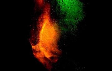
A light-sheet microscope 3D image displays the convergence of dopaminergic transmitter inputs from the ventral tegmental area (green), a midbrain structure associated with reward and motivation, and glutamatergic inputs from the ventral hippocampus (red), an area deep within the brain that helps with navigation, onto the nucleus accumbens. [Image: Kanghoon Jung]
Animals often navigate their surroundings with a specific goal in mind, such as seeking out food, water or shelter. They form spatial memories tied to these goals, that are stored in the brain and later retrieved to guide future navigation.
Researchers in the United States have successfully reactivated a spatial memory in mice, causing the mice to attempt to shelter when no shelter is actually present (Nat. Neurosci., doi: 10.1038/s41593-024-01770-9). They used a light-activated gene-expression technique called Cal-Light, which enabled them to switch on the expression of the genes associated with shelter memory neurons.
The researchers believe that the work could provide a foundation for reactivating or engineering memory circuits in people with neurodegenerative disorders like Alzheimer’s disease.
Uncovering neural circuitry
Kanghoon Jung, a postdoctoral research fellow in the lab of Hyung-Bae Kwon at Johns Hopkins University School of Medicine, studies the neural circuitry underlying “where-to-go” decisions. The brain structure serving these goal-oriented spatial memories must receive information about the spatial context and an animal’s current needs to provide the necessary navigation toward a goal. However, causal links between specific neuronal populations and goal-directed navigation are still poorly defined.
To explore these connections, Jung and his colleagues imaged and stimulated neurons in two areas of the brain—the nucleus accumbens, involved in motivation and reward, and the dorsal periaqueductal gray, associated with escape responses—to explore their role in the formation of spatial memories. They employed Cal-Light, an optogenetic system developed by Kwon in 2017 that allows selective tagging and subsequent controlling over neuronal populations in a user-defined time window using light.
“By illuminating the nucleus accumbens with blue light when the mouse was in the shelter, we tagged neurons associated with shelter-seeking,” said Jung. “Later, we reactivated these neurons, along with artificial activation of the escape brain circuit, causing mice to seek the place previously associated with the shelter, even without an active threat or the shelter present.”
The results suggest that a memory module in the nucleus accumbens, where spatial information and safety dopamine signals converge, is responsible for encoding spatial goal locations and facilitating goal-directed navigation.
Role of dopamine
The researchers also used fiber photometry coupled with genetically encoded biosensors to reveal how dopamine signals associated with safety correlate with other neuronal activities during shelter experiences. Notably, dopamine is released into the nucleus accumbens in response to rewarding stimuli.
The results suggest that a memory module in the nucleus accumbens, where spatial information and safety dopamine signals converge, is responsible for encoding spatial goal locations and facilitating goal-directed navigation. Also, the work reveals the role of dopamine in the formation of memory-linked neural circuits.
“Real-world applications could involve developing novel dopamine-mediated treatments for memory disorders like Alzheimer’s disease,” said Jung. “The technique of tagging specific memory-associated cells also opens possibilities to identify relevant cell types and proteins essential for memory formation, which may lead to more effective treatments for neurodegenerative diseases.”