
![]()
Yoan Simon of Arizona State University, USA, was one of the principal investigators behind new work that used lasers, fluorescent mechanophores and high-speed photography to illuminate how energy is dissipated in high-speed impacts. [Image: Y. Simon]
Understanding what happens in a high-velocity impact is important across a range of disciplines and scales, from meteorite strikes in planetary studies to microscopic failure assessment in materials science. But as might be expected from something as disorderly as an impulsive impact, the science of such events can also be a bit messy—with different research communities understanding the relevant phenomena in different ways.
A multi-institution US research team has combined some creative chemistry, fluorescence microscopy and laser-driven microballistics to gain a clearer view of just what happens in a high-strain-rate impact, and to tease out the relative contribution of different kinds of energy dissipation within the target (Nat. Commun., doi: 10.1038/s41467-024-52663-1). At the heart of the method are tiny beacons called mechanophores—molecules that, under a sufficiently large mechanical force, light up with a long-lasting fluorescent signal, helping to provide a high-resolution 3D map of the deformation.
The issue: characterizing energy dissipation
One reason impulsive impacts are so challenging to understand is that the energy of the impactor tends to be dissipated in different ways in the target. Part of the energy might be soaked up by attenuation of an acoustic wave in the material upon impact—an issue for the shockwave-physics community and commonly a preoccupation for researchers in Earth and planetary science. On the other hand, part will also be taken up by an inelastic failure mechanism such as plastic deformation, the focus of materials scientists and engineers.
According to the authors behind the new research, it’s been difficult to reconcile these views, as the approaches for assessing damage mechanisms tend to stop at the material’s surface and to take place only after the impact has taken place.
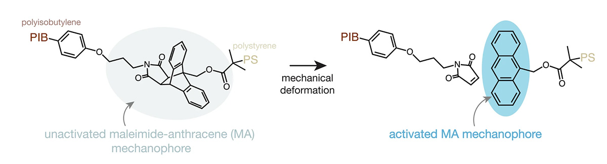
When the polymer material is subjected to sufficiently intense mechanical deformation, the mechanophore developed by the research team breaks apart, releasing a fluorescence signal. [Image: P.J. Centellas et al., Nat. Commun., doi: 10.1038/s41467-024-52663-1; CC-BY 4.0]
To get at a deeper view, the team behind the new work—led by Edwin Chan at the US National Institute of Standards and Technology (NIST) and Yoan Simon at Arizona State University (ASU), USA—turned to a maleimide-anthracene (MA) mechanophore as a sort of undercover spy on the deformation process. When subjected to a sufficient impulsive force, the MA mechanophore will break into two different species, one of which will glow fluorescently under UV-A irradiation. Even better, the activated sites will remain fluorescent for up to two days.
The researchers developed a method to chemically incorporate the MA mechanophores into a polyisobutylene-b-polystyrene (PIB-b-PS) diblock copolymer, and to draw the mechanophore-impregnated copolymer out into a thin film. The result was a “self-reporting” material that would light up like the proverbial Christmas tree when dealt a sufficient shock after a high-strain-rate impact.
Laser-driven microballistics
With the mechanophore-doped polymer in hand, the team next subjected it to some very tiny but very nasty impacts. For this part of the experiment, they used a technique called laser-induced projectile impact testing (LIPIT).
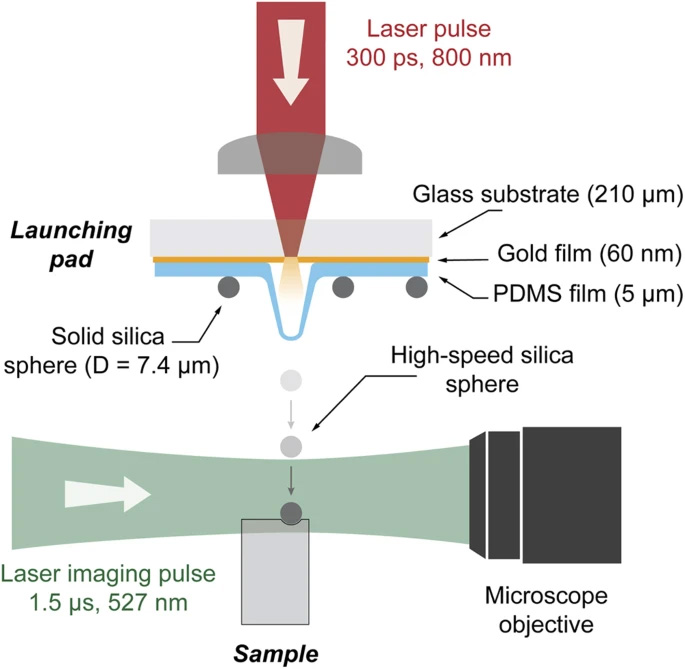
A typical setup for laser-induced projectile impact testing (LIPIT). [Image: D. Veysset et al., Sci. Rep., doi: 10.1038/srep25577 (2016); CC-BY 4.0]
In a typical LIPIT trial, a laser excitation pulse is focused on a substrate containing an ablation material such as a thin gold film, with silica or other microparticles embedded in a layer of elastomer film such as polydimethylsiloxane (PDMS). When the laser pulse hits the gold film, ablation of the material leads to the creation of a plasma that expands and drives the microparticle forward like a bullet, at speeds of up to 1 km/s. The actual speed can be controlled by varying the thickness of the elastomer film layer.
The researchers behind the new work used such a setup to fire individual 20-µm-diameter silica microparticles toward the MA-mechanophore-containing copolymer film. They filmed the trajectory and impact of the blitzing particles using an ultrafast camera, capturing frames with an exposure time of 30 ns each, and with 150 to 450 ns between frames depending on impact velocity. The actual impact velocities in the experiments ranged from 100 to 520 m/s.
The ultrafast photography allowed the team to approximate the penetration strain rate in the material on impact—which turned out to be very high (on the order of 108 s–1). In addition, from the rebound velocity of the particles, the researchers could estimate the kinetic energy loss, and thereby back out the amount of energy dissipated in the film for a given impact velocity.
Mechanophores and Mach cones
Next, the team took a close look at the damaged material to try to separate out the energy dissipation mechanisms at play. They started by using atomic-force microscopy (AFM) to measure the surface profile of the deformed material. They found that it included shallow craters with material pile-up around the rims, a classic pattern indicating energy dissipation by plastic deformation.
Next, the team put its MA mechanophores to work, using fluorescence microscopy to visualize where the impact had sufficiently disturbed the molecular beacons to break them apart and trigger the fluorescence response. As expected, they found a strong signal in the area of the crater and its perimeter. But interestingly, they also found that for the trials with faster impact velocities, the mechanophores had been fluorescently activated well below the surface of the impacted site as well, in a zone with the shape of a cone.
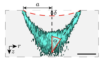
The team found that the fluorescence signal from the MA mechanophores was arrayed in the shape of a Mach cone, below the level of the actual impact damage (dashed red line)—an indication that part of the impact energy was being dissipated by acoustic-wave attenuation rather than plastic deformation. [Image: P.J. Centellas et al., Nat. Commun., doi: 10.1038/s41467-024-52663-1; CC-BY 4.0]
According to the authors, these subsurface, mechanophore-activated cones “bear a striking resemblance” to so-called Mach cones, the conical pressure waves formed in the surrounding medium by a body traveling at greater than the speed of sound. Finite-element computational modeling of the process backed up the notion that the cone-shaped structures were related to the dissipation of hypersonic shear waves within the material.
Toward a deeper understanding
The researchers believe their technique can help discriminate the relative importance of acoustic-wave attenuation and plastic deformation in high-strain-rate impacts—and thus that it “bridges viewpoints from both impact physics and shockwave physics communities.” They go on to suggest that that extending the platform to other mechanophore-functionalized materials might yield insights into a range of impulsive events, including mild traumatic brain injuries, cold spray additive manufacturing and hypervelocity impacts in space.
ASU’s Yoan Simon, a co-PI on the work, also sees potential applications in environmental protection. In a press release accompanying the work, he noted that a key way to reduce the need for new plastics in the environment is to prolong the lifespan of existing ones. An important prerequisite to that, Simon suggested in the release, is to better understand what happens when a material responds to shocks—an understanding that he believes the team’s method could afford. “What’s really novel about this study,” he said, “is that it provides us with a unique tool to look at what happens deep inside the material.”
In addition to researchers at NIST and ASU, the study also included contributions from team members at the University of Southern Mississippi, USA, the US Army Engineer Research and Development Center, and Rensselaer Polytechnic Institute, USA.