
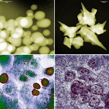
Spherical and star-shaped gold nanoparticles (top) and colon cancer cells after about five hours of exposure to them (bottom, respectively). The photo in the bottom left corner shows that, despite the small size of the spherical nanoparticles, the cancer cells survived. [Image: IFJ PAN]
Researchers in Poland have discovered that the shape, rather than the size, of gold nanoparticles matters most when it comes to killing cancer cells (Small, doi: 10.1002/smll.202400778). They performed live cell imaging with holotomography—a real-time, label-free laser technique that measures the 3D refractive index of a sample—finding that star-shaped nanoparticles perforate cell membranes and trigger cell death.
The work adds to our understanding of the mechanical interaction between cancer cells and gold nanoparticles, which may pave the way for the development of more effective, better-targeted therapies.
Advantages of holotomography
Live cell imaging allows scientists to observe the inner workings of cells without interfering with their natural physiology. Holotomography is a relatively new live cell imaging technique, which measures the 3D refractive index distribution within the cell in real time, noninvasively and without the addition of fluorophores, a fluorescent chemical compound used in microscopy to enhance visuals.
The researchers wanted to test whether the common assumption of “smaller kills faster” rang true for different shapes of gold nanoparticles. With the Nanolive 3D CX-A holotomographic microscope, they were able to visualize specific cell compartments and nanoparticle accumulation sites with little to no phototoxicity.
“Unlike other high-resolution microscopy techniques, holotomography does not require the preparation of samples or the introduction of any foreign substances into the cells,” said study author Joanna Depciuch-Czarny, Institute of Nuclear Physics of the Polish Academy of Sciences, in a press release accompanying the research. “The interactions of gold nanoparticles with cancer cells could therefore be observed directly in the incubator, where the latter were cultured, in an undisturbed environment, what’s more with nanometric resolution, from all sides simultaneously and practically in real time.”
Creating a model
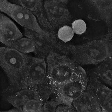
Interaction with small spherical gold nanoparticles did not change the morphology of colon cancer cells, and the cells are still able to divide. [Image: IFJ PAN]
Depciuch-Czarny and her colleagues synthesized three types of gold nanoparticles: sphere-like shapes with an average size of 10 nm, star-like shapes with an average size of 244 nm and rod-like shapes that measured approximately 45 nm × 10 nm. Experiments involved culturing the nanoparticles with two different glioblastoma cell lines and one colon cancer cell line with images taken after 10, 180 and 285 minutes.
While the smaller spherical nanoparticles easily entered the cancer cells, the cells were able to recover and keep dividing. The larger star-shaped nanoparticles punctured membranes as they aggregated inside the cell, causing irreparable damage and eventually leading to apoptosis. Based on these data, the researchers built a theoretical model of the absorption dynamics of gold nanoparticles by cells.
“The final result is a differential equation into which suitably processed parameters can be substituted―for the time being only describing the shape and size of nanoparticles―to quickly determine how the uptake of the analyzed particles by cancer cells will proceed over a given period of time,” says study author Pawel Jakubczyk at the University of Rzeszow. “Any scientist can already use our model at the design stage of their own research to instantly narrow down the number of nanoparticle variants requiring experimental verification.”