

Rembrandt van Rijn, Militia Company of District II under the Command of Captain Frans Banninck Cocq, known as The Night Watch (1642); Rijksmuseum, Amsterdam, on loan from the City of Amsterdam. Researchers have used a combination of X-ray imaging and tomography techniques to shed new life on how Rembrandt created the celebrated, massive painting. [Image: Rijksmuseum media gallery]
It’s one of the most recognizable images from the Dutch Golden Age (1588-1672 C.E.): the vast oil canvas by Rembrandt van Rijn commonly called The Night Watch. More than 15.8 m2 (12×14.5 ft) in size, the painting almost jumps off the wall with energy and life. And even today, art historians and conservators continue to study The Night Watch for clues on how Rembrandt managed the accomplishment.
That quest lies behind “Operation Night Watch,” a multiyear research and conservation project begun in July 2019 at the Rijksmuseum, Amsterdam, Netherlands, to get to the bottom the masterwork, both literally and figuratively. In the project’s latest finding—based on synchrotron X-ray fluorescence (XRF) imaging and a computational-imaging technique known as ptychographic X-ray computed tomography (PXCT)—scientists have drawn an intriguing inference about the picture’s creation (Sci. Adv., doi: 10.1126/sciadv.adj9394). Faced with the display and preservation challenges of the huge artwork, the canny Rembrandt may have opted to wash the canvas with an innovative lead-based, moisture-protective coating before starting work.
The museum says such lead-based impregnation “has never before been observed with Rembrandt or his contemporaries,” and indicates the artist’s openness to new ideas. The scientists, meanwhile, note that the study marks the first use of these combined synchrotron-based techniques on a historic paint microsample. And they think the combination of XRF imaging and ptychography could become “a valuable tool” in such studies as both synchrotron brightness and computer capabilities continue to improve.
Big painting, big challenges
In painting as large a canvas as The Night Watch—officially titled Militia Company of District II under the Command of Captain Frans Banninck Cocq—Rembrandt knew he faced some practical problems. One was simply the big painting’s uncommon weight. That factor, art historians believe, may have put the artist on a search for lighter ground-coating materials than the combination of iron-rich red pigments and lead-white paint he used in earlier canvases.
In painting The Night Watch, Rembrandt faced some practical problems—related both to the painting’s size and where it would be displayed.
Another challenge, though, related to the spot where the painting would actually be displayed—the outer wall of the great hall of the Kloveniersdoelen, a “musketeer’s” shooting range in Amsterdam. That relatively humid location posed a threat to the painting’s long-term staying power, as the animal-glue sizing layers typically used in 17th-century canvas preparation afforded inadequate protection from moisture and mold.
Previous studies, using techniques such as polarized-light microscopy, energy-dispersive X-ray spectroscopy and small-angle X-ray diffraction, had already revealed that the painter had departed from his previous practice and selected a different, lighter quartz-clay ground material for The Night Watch. As part of its conservation and restoration effort, the Operation Night Watch team wanted to get a better view of the 3D morphology and condition of that quartz-clay ground.
Scalpel, stereomicroscope and synchrotron
To do so, the team—which included researchers from the Rijksmuseum, the University of Amsterdam, the University of Antwerp and Utrecht University—used a scalpel and a stereomicroscope to take a sub-millimeter-square sample from the huge canvas. The sample was gathered from a seam on the picture where two canvas strips were joined. That sampling spot made the ground layer particularly accessible owing to the loss of some of the overlying paint over time.
The scientists then took the sample to the PETRA III synchrotron radiation source at the Deutsches Elektronen-Synchrotron (DESY) facility in Hamburg, Germany. At DESY, the tiny paint chip was bombarded by monochromatic, 17-kEV X-rays delivered in a 200×300-nm beam, with the sample on a rotating stage to allow a 360-degree scan. The researchers trained the X-rays on a 55×160-µm area of the sample fragment, and collected XRF data using two secondary X-ray detectors. They also gathered far-field diffraction data for PXCT with a hybrid pixel detector placed 8 m from the sample.
Combining techniques
At DESY, the tiny paint chip was bombarded by monochromatic, 17-kEV X-rays delivered in a 200×300-nm beam.
XRF works by shining hard-X-ray light on a sample to excite inner-core electrons of specific atoms, and then collecting secondary X-ray energy re-emitted at different frequencies when the electrons relax back to the ground state. The characteristic frequencies of that secondary radiation can be tied to the abundance of certain heavy elements such as iron, calcium and lead (see “The Art of Discovery,” OPN, January 2023).
In PXCT, meanwhile, analysis of a multitude of far-field diffraction patterns enables a 3D model of the electron density in a sample to be computationally built up—and also allows visualization of low-atomic-number elements that XRF would miss. Combining XRF and PXCT, the authors note, has proved useful in studies of biological samples and chemical catalysts.
A protective wash of lead?
Applied to The Night Watch, the correlated techniques yielded up some surprises. The biggest was that the fragment of ground material imaged and mapped by PXCT contained copious amounts of lead as revealed by XRF. A deeper dive into the ptychographic signal revealed that the lead was concentrated in the lowest part of the sample—that is, the part closest to the canvas.
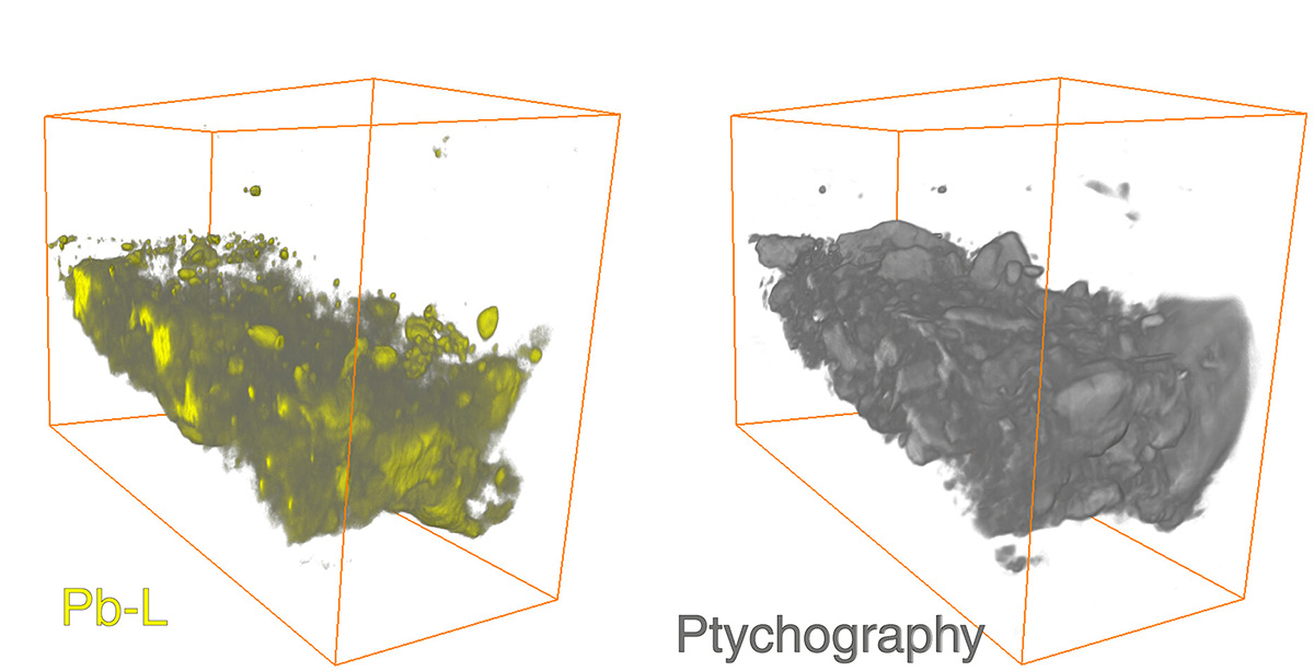
Studying the paint sample with XRF and 3D ptychographic tomography, the researchers found a surprising abundance of lead in the quartz-clay ground material—with the lead particularly concentrated on the part of the ground closest to the canvas. [Image: F.T.H. Broers et al., Sci. Adv. 9, adj9394 (2023), doi: 10.1126/sciadv.adj9394; CC-BY 4.0]
The abundance of lead constituted an unexpected finding for the lightweight quartz-clay ground material in the painting. That discovery, however, chimed with larger, noninvasive macro-XRF element-distribution maps of the painting as a whole. Those maps had shown copious amounts of lead in The Night Watch, much of it seemingly applied in large, circular brushstrokes.
The presence of the lead had previously been partly attributed to the use of lead-containing white pigments in some areas of the painting, to depict features such as faces and the era’s evocative ruffled collars. But the lead mapped in the darker regions was something of a mystery—as was the presence of tiny “lead soap” protrusions across the entire canvas.
The researchers hypothesize that the abundance of lead in The Night Watch could instead relate to goings-on well below the surface. Before applying the quartz-clay ground material, they suggest, Rembrandt—mindful of the painting’s ultimate location in a damp shooting-club hall—may have washed the canvas in a lead-containing oil layer, to provide better protection from the elements. The idea was in the air at the time; a 17th-century treatise on art techniques by Théodore de Mayerne, for example, suggested impregnation with lead-rich oil as an alternative to the inferior animal-glue sizings then commonly used for protection.
New conservation tool
A press release from the University of Amsterdam said the discovery “underlines Rembrandt’s inventive way of working, in which he did not shy away from using new techniques.” In the paper, the researchers likewise highlight the work’s importance both in revealing aspects of the artist’s creative process and in providing the detailed understanding needed for practical work toward restoration and reconstruction.
The scientists also believe that the results of their study—the first, to their knowledge, to combine synchrotron radiation–based correlated XRF and ptychographic nanotomography on a historic paint micro-sample—suggest that the technique could be useful in other, future conservation work, particularly to provide ground truth for interpreting noninvasive techniques such as macro-XRF mapping. The combination, the team believes, could prove “a valuable tool for future cultural-heritage studies.”